
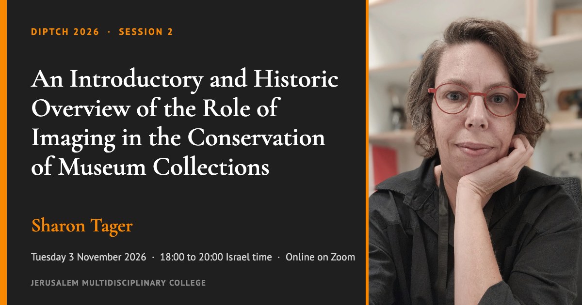
Sharon Tager, Head of Conservation at the Israel Museum, Jerusalem, traced how conservation grew from a craft into a science, and how imaging became one of its main tools for making decisions. Start with the key takeaways, then open any section below for the history, the case studies from the Israel Museum, the techniques and terms, and links for going further.
Key takeaways
- Conservation decisions draw on several kinds of knowledge at once: history and context, ethics, science and technology, and the conservator’s hands-on skill.
- Imaging serves three main purposes in conservation: documentation, identification of materials and techniques, and visualization.
- Examination starts without touching the object: the eye, magnification, ultraviolet, infrared, multiband imaging and X-ray come first. Conservators take a physical sample only if questions remain.
- Imaging supports a decision; it does not make it. Its results are read together with the object’s history, its condition and the curator’s view.
- Treatments use reversible materials, so that future generations can revisit today’s choices as knowledge changes.
How the session ran
Sharon presented in two parts with a break, and invited questions throughout rather than only at the end. Moshe Caine, who documented one of the case studies with photogrammetry, joined the discussion. The talk moved from the history of the profession to current practice in the Israel Museum’s conservation department.
How conservation became a science
Before the nineteenth century, restorers routinely repainted, regilded, cut down or added to damaged objects; the idea of the original as something to preserve came later. Sharon traced the turn to the Victorian critic John Ruskin, who argued against drastic alteration of monuments.
Later, science entered the museum with the chemist Friedrich Rathgen, the first head of the chemical laboratory founded at the Royal Museums in Berlin in 1888, today the Rathgen-Forschungslabor.
Researchers used X-rays on cultural objects as early as 1896. In the United States, the Fogg Museum at Harvard became a center of technical study: its director Edward Forbes began a pigment collection to understand how artists’ materials were made and used. At the Metropolitan Museum of Art, James J. Rorimer published Ultra-Violet Rays and Their Use in the Examination of Works of Art in 1931; artists painted its color plates of fluorescence by hand.
Meanwhile, military and medical technology drove much of the progress. In the late 1960s, J. R. J. van Asperen de Boer developed infrared reflectography in the Netherlands. The method shows the underdrawing beneath the paint, and his early infrared images included the Ghent Altarpiece.
The cleaning controversy around the National Gallery in London, from 1947 to 1963, set the gallery’s science-led approach, carried out by the conservator Helmut Ruhemann, against critics such as Cesare Brandi and Ernst Gombrich, who argued that paintings were being over-cleaned. As a result, the debate pushed the field toward documented, transparent, evidence-based decisions.
Today the field is shifting again, toward a balance between scientific evidence and ethical, historical and cultural judgment.
How imaging supports decisions
- Three purposes. Documentation records an object’s condition before, during and after treatment. Identification reveals materials and techniques, often without a sample. Finally, visualization makes the findings visible to researchers, curators and the public.
- Noncontact first. The workflow begins with ordinary light and magnification, then ultraviolet, infrared, multiband imaging and X-ray where relevant. Conservators consider physical sampling only when these steps leave questions unanswered. Keyword: Non-invasive documentation.
- Shared decisions. The conservator is responsible for treatment materials and technical execution. However, the conservator decides together with the curator on varnish removal, how far to clean, which later additions to keep and the style of restoration. Display questions, such as light levels for drawings, watercolors and textiles, are where their priorities most often clash.
- Reversibility. Conservators can change earlier reconstructions when new archaeological or historical research supports a different arrangement, and new reconstructions for an upcoming Israel Museum exhibition on early synagogues will use reversible materials. Keyword: Reversibility.
- Reading the results. A pigment distribution map shows where an element is found, not the real color of the paint. For this reason, conservators interpret imaging alongside historical research, the object’s condition and their own expertise. Keyword: Authenticity and integrity.
Case studies from the Israel Museum
- Raking light. Photographs of a stone stela in raking light revealed inscriptions on its back that had not been recorded. The conservation department’s photographer, a graduate of the Photographic Communication program at JMC, took them.
- Ultraviolet fluorescence. On a panel painting from the Fayum in Roman Egypt, the characteristic fluorescence of madder lake identified the dye in the highlights without taking a sample.
- Canvas weave. For a research project and exhibition on Amedeo Modigliani’s materials and techniques, the museum sent the X-ray of its Modigliani, Jeanne Hébuterne, Seated (1918), to a specialist in automatic thread counting. The weave pattern works like a fingerprint and linked the painting to other works made from the same roll of canvas, which helps group and date them. Read more: Interpreting Canvas Weave Matches (ArtMatters, 2013).
- Pigment maps. Sharon is researching the materials and techniques of the Israeli painter Reuven Rubin, turning X-ray fluorescence data into pigment distribution maps like the false color image of his self-portrait on her page.
- A hidden change. Imaging of a nineteenth century Persian portrait of Fath-Ali Shah showed that the painter first gave him a long sword and later painted it over with a dagger.
- Monitoring change. Many of Georgia O’Keeffe’s oil paintings develop tiny protrusions of lead soaps. As a result, conservators monitor them by combining photometric stereo under UV-induced fluorescence with X-ray fluorescence. Read more: Diagnosing “Art Acne” in Georgia O’Keeffe’s Paintings (Northwestern University).
- Dead Sea Scrolls. Conservators compare infrared photographs of scroll fragments from 1949 with new infrared images to detect changes in size, deformation, ink loss and new cracks. Images of the scrolls are available in the Leon Levy Dead Sea Scrolls Digital Library.
- A fragile ivory head. A burnt ivory head from the Late Bronze Age was too delicate to handle while planning its display. Moshe Caine documented it with photogrammetry, and a 3D print made at the same weight as the original let conservators and art handlers test the mount before touching the object. The head and the ivory harp piece were both excavated at Tel Miqne, in different years. The object was shown with its reconstruction in the Israel Museum’s exhibition Feast, curated by Nurith Goshen, with research by Baruch Brandl. Read more: The glory of ancient feasts is being revived at the Israel Museum (The Jerusalem Post, 2023), and, in Hebrew, a short text with an image of the harp in Koch (ed.), In Centro, Collected Papers VI: Feast (2026), p. 130. Keywords: Photogrammetry, Digital duplication and 3D printing.
- Public access. Closer to Van Eyck lets anyone explore the Ghent Altarpiece in macro photography, infrared reflectography and X-radiography, before, during and after its treatment. Sharon presented it as a model for sharing conservation imaging with the public.
More objects from the talk
- Elijah’s chair. Bezalel School, Jerusalem, 1916 to 1925, designed by Ze’ev Raban. The museum’s online record does not yet show the new photograph taken after conservation or the list of the chair’s materials. See the Israel Museum collection record.
- The statue of Hermes from Beth She’an. A marble statue from the Severan Theater at Beth She’an, restored and displayed at the Israel Museum. Further reading: Tali Sharvit, “Three Marble Statues from the Severan Theater,” chapter 10 in G. Mazor and W. Atrash, Bet She’an III/2 (IAA Reports 58/2), 2015.
Techniques and terms
- X-radiography. X-rays pass through the object and show what lies beneath the surface: earlier compositions, the structure of a panel or canvas, and internal repairs. Keyword: X-radiography.
- Ultraviolet fluorescence. Under ultraviolet light some materials glow in a characteristic way, which reveals varnish layers, retouching and certain pigments and dyes. Keyword: Ultraviolet fluorescence.
- Infrared reflectography. Infrared passes through many paint layers and shows the underdrawing and changes in the composition. Keyword: Infrared reflectography.
- Multiband imaging. A series of images of the same object across ultraviolet, visible and infrared light, compared to tell materials apart. Keyword: Multispectral imaging.
- Raking light. Light from a low angle across the surface brings out texture, tool marks and faint inscriptions. Keyword: Raking light.
- X-ray fluorescence mapping. Maps where chemical elements are found across a surface, which points to the pigments used; the colors of the map are not the colors of the paint. Keyword: X-ray fluorescence mapping.
- Canvas weave analysis. Thread counts measured from X-rays give each canvas a pattern; matching patterns show that paintings were cut from the same roll. Keyword: Canvas weave analysis.
- Photometric stereo. Images taken with light from several directions are combined to calculate the shape of the surface, which makes it possible to measure small changes over time. Keyword: Photometric stereo.
AI in conservation
On AI, Sharon explained that most conservators are conservative by nature, though not all. At the Israel Museum, for now, the AI applications being adopted are mainly about collecting and analyzing data, particularly in preventive conservation: for example, the vast data from relative humidity, temperature and light exposure measurements. For classic conservation and restoration, the museum has not yet found a direct link between AI and the work.
Read more on research into AI for the restoration of paintings: Have a damaged painting? Restore it in just hours with an AI-generated “mask” (MIT News, 2025).
Questions to carry forward
- Who decides what stays and what is removed, and how should a conservator and a curator resolve a disagreement?
- When does a question justify taking a physical sample from an object?
- Imaging produces more data than ever. How should it be stored, interpreted and shared with the public and with future generations?
- Is a digital copy a form of conservation, or does the physical object still need exactly the same care?
- Where should AI fit into conservation decisions, and how can its results stay transparent?
- The thread continues next week with Shai Halevi on advanced imaging technologies in archaeology, and on 22 December with Graham Diprose on photography past, present and future.
Keywords for this session: X-radiography · Ultraviolet fluorescence · Infrared reflectography · Multispectral imaging · Raking light · X-ray fluorescence mapping · Canvas weave analysis · Photometric stereo · Photogrammetry · Digital duplication and 3D printing · Non-invasive documentation · Reversibility · Authenticity and integrity